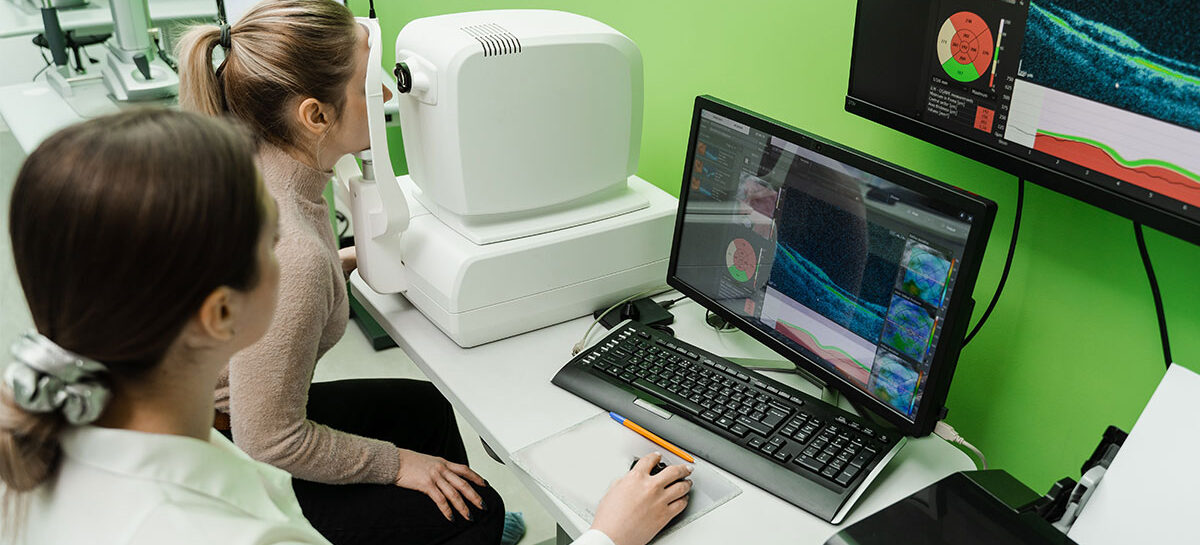
Optical Coherence Tomography procedure used in Cardiology

Summary
Optical Coherence Tomography or OCT is a minimally-invasive procedure done to get a better idea of blockages in coronary arteries. Compared to other options such as coronary angiography and intravascular ultrasound, OCT offers many advantages. This is what is making the procedure popular although it has not reached the stage of mass adoption. It is used along with coronary angiography most of the time. In this article, we will learn more about OCT.
Introduction to PCI
Atherosclerosis is a condition in which cholesterol, calcium and some other material found in blood flowing through arteries start depositing on the inner walls of the artery. Over time, these deposits narrow down the artery and block blood-flow, which causes various complications. Further, the deposits harden into what is called plaque. The plaque can break off into pieces which can tear the artery wall, causing blood to clot there and cause new complications. Atherosclerosis can happen in arteries anywhere in the body, but the consequences are more severe when they happen in coronary arteries – or arteries of the heart, and this includes heart failure and heart attack.
Percutaneous Coronary Intervention (PCI) is a minimally-invasive procedure aimed at opening out coronary arteries that have been blocked due to atherosclerosis. In the past, PCI meant ‘coronary angioplasty with stenting’, as coronary angiography was the only technique used for PCI imaging. However, today, there are other options available too, as mentioned below.
Also Read: Techniques superior to Angiogram to identify heart blocks
PCI Imaging Options
An important aspect of treating atherosclerosis is understanding the blockage correctly. What kind of material is there (lipid-rich plaque, fibrotic plaque, vulnerable plaque or calcified lesion), what is the length and depth of the lesion (lesion means abnormal growth, but in this case, it is referring to the blockage), the diameter of the lumen or blood-vessel concerned, etc.
Getting all these details right is important to decide specifications of the stent that will be inserted at the blockage, after balloon angioplasty is done. Balloon angioplasty involves inflating a balloon at the site of blockage. This will push the deposit material against the wall and clear the blockage. The stent (a wire-mesh basically, that looks like a spring) is inserted to prevent the deposits from growing again and creating a repeat blockage.
Needless to say, the quality of imaging (photos or videos captured of the blockage) is critical for the right intervention, or to ensure the right stent is inserted.
Coronary Angiography (CA)
In this procedure, a catheter or a narrow, thin and flexible tube is inserted into the groin or arm, after making an incision (cut there). The catheter is then threaded all the way to the heart. A contrast medium (basically a liquid dye) is flushed into the catheter and hence the blood-vessels of the heart. A series of X-ray (fluoroscopy) images are taken and these map the flow of the dye which is visible in the X-ray. This way, the number, location and size of the blockages can be determined. Once the doctor is satisfied, the catheter is pulled back and out of the body.
Intravascular Ultrasound (IVUS)
Intravascular Ultrasound (IVUS) is a better option than CA. Similar to CA, an incision is made in the groin and a catheter inserted there. The catheter is threaded all the way to the heart and up to the farthest point that needs to be photographed. The catheter also carries a guide-wire with an ultrasound probe at the tip. The probe will emit ultrasound waves, and the reflected waves from the inside of the artery are captured by the probe as echoes and relayed to a computer monitor kept in the procedure room.
The guide-wire is held in place and the probe is pulled out steadily using motorized control (process called pullback). In the process, it continuously relays images to the screen. IVUS is a better option than CA as it can reveal the percentage of stenosis (blockage), the type of plaque and whether there was a restenosis (repeat blockage) at the site that was recently cleared.
OCT – Optical Coherence Tomography
- as discussed below.
What is OCT and how is it done?
OCT is the most sophisticated imaging technique used in PCI, far better than CA or IVUS. OCT uses infrared light and the reflected light is captured to create an image of the inside of the artery. The images are high-resolution (almost 10x that of images obtained using IVUS), so the doctors have a better idea of the blockage, the nature of plaque etc.
In the initial days, OCT was used to obtain a clear picture of the back of the eye. This was required to treat vision problems like diabetic-retinopathy and glaucoma. Pretty soon, OCT was being adopted in cardiology as a superior option for PCI Imaging.
Today, OCT is used as an adjuvant, or additional tool, to
- assess the morphology and composition of atherosclerotic plaques
- guide and optimize the coronary angioplasty procedure, especially with regard to stenting (both in pre-stenting and post-stenting tasks)
OCT is increasingly used along with CA. This is known to be a better option than using CA alone, or OCT alone. It can detect the extent of fatty deposits, can detect the presence of clots, and take precise measurements required for placing the right stent.
In an OCT procedure, the catheter is inserted as described in the other procedures. The catheter carries a single optical fibre in which near infrared light is emitted. The light emitted gets reflected by the walls of the artery and any structure present there such as plaque deposits or a clot. Some of the light also gets scattered. The scattered light, also called glare, is of no use, and is filtered out by the OCT.
The reflected light is of interest to us. Even a tiny amount of reflected light can give us several and useful images. The time-delay in reflection and the signal strength of the reflected light are captured and relayed to a computer monitor that will convert the reflected light-signals into powerful or high-resolution images. The optic fibre is pulled back along the length of the coronary artery in a motorized fashion (called pullback), which helps scan the areas of interest in the artery.
Also Read: The Role of a Cardiac Electrophysiologist
Benefits or Advantages of OCT over other options
Compared to IVUS, OCT offers several benefits or advantages such as:
- Better image resolution (10x compared to IVUS)
- Precise estimation of calcified lesions
- In cases of stent failure, OCT provides better assessment of both acute and chronic failures
- Better identification of minor lesions such as tears and intimal erosions
- assessing the long-term safety and effectiveness of the implanted stent
In fact, OCT provides very detailed information about the blockage such as:
- Edge dissection (disruptions in the artery’s inner surface at the edge of the stent)
- Tissue prolapse (the arterial wall tissue protrudes into the gaps in the stent after the stent is placed)
- Thrombi or clots, and
- Incomplete stent apposition or stent malapposition, which is lack of proper contact between the stent and artery wall required for optimal stent deployment
These details are important for successfully reducing stent-related complications and MACE or major adverse cardiovascular events (such as non-fatal stroke, non-fatal heart-attack, heart-failure and cardiac death).
Kauvery Hospital is globally known for its multidisciplinary services at all its Centers of Excellence, and for its comprehensive, Avant-Grade technology, especially in diagnostics and remedial care in heart diseases, transplantation, vascular and neurosciences medicine. Located in the heart of Trichy (Tennur, Royal Road and Alexandria Road (Cantonment), Chennai (Alwarpet & Vadapalani), Hosur, Salem, Tirunelveli and Bengaluru, the hospital also renders adult and pediatric trauma care.
Chennai Alwarpet – 044 4000 6000 • Chennai Vadapalani – 044 4000 6000 • Trichy – Cantonment – 0431 4077777 • Trichy – Heartcity – 0431 4003500 • Trichy – Tennur – 0431 4022555 • Hosur – 04344 272727 • Salem – 0427 2677777 • Tirunelveli – 0462 4006000 • Bengaluru – 080 6801 6801
- May 03, 2024
