
The O-ARM Surgical Imaging System – Enhanced Precision, Versatility and Safety
 Summary
The O-ARM surgical imaging system is an advanced imaging tool designed to improve precision during spinal and cranial surgeries. It creates real-time 2D and 3D scans of the patient using continuous X-rays during surgery. This helps surgeons navigate complex and sensitive structures with accuracy. The O-ARM machine is shaped like a C-arm that forms a complete ring around the patient once it is in position. The ring rotates and provides 360-degree visualization. Unique features of the O-ARM include multiple fields of view, low-dose radiation mode, and stereotactic mode – The O-ARM is truly the future of high-tech spine surgery. Fully optimized for AI and VR integration, the O-ARM eliminates the need for separate pre-op and post-op scans. This saves critical time and streamlines the process. The O-ARM is useful in procedures like spinal fusion, tumor removal, joint replacement and spinal fracture repair. The system offers better outcomes, smaller incisions and faster recovery. The O-ARM represents a leap forward in surgical accuracy, safety and efficiency.
What is the O-ARM Surgical Imaging System?
The O-ARM surgical imaging system is a state-of-the-art visualization tool used during brain and spinal surgery. The system uses continuous X-rays from multiple angles to construct a real-time 3D model of the patient’s anatomy during surgical procedures. The system can also provide detailed 2D visualization, depending on what the surgeon requires. The O-ARM system allows the surgeon to visualize the surgical area, as well as guide surgical instruments with unprecedented precision. The O-ARM technology is based on portable CT imaging technology. It is a unique, patented technology that integrates next-gen anatomical visualization with surgical robotics. The O-ARM system is also fully mobile, making it useful in adjusting patient positioning during surgery. The system also has the ability to store specific viewing angles, allowing the surgeon to switch quickly between key viewing angles in real time. The O-ARM is designed to provide accurate visualization with low X-ray exposure, minimizing potential radiation damage for both patients and surgical staff. It follows the principle of ALARA – As Low As Reasonably Achievable. This is a guiding principle in radiation safety, emphasizing that radiation exposure needs to be kept as low as possible, while still achieving the desired effect – Even below regulatory limits if possible. O-ARM is also optimized for virtual reality and AI integration – This will make it even more convenient and intuitive for surgeons to use.
How does the O-ARM work?
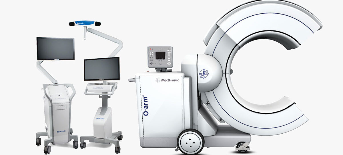
The O-ARM machine is shaped like a C arm attached to an X-ray console on wheels. It has another telescopic arc which can be extended out from the C to form a complete ring around the patient on the surgical table, reminiscent of an MRI machine. The ring rotates around the central axis of the flatbed the patient’s body is positioned on. This gives the surgeon 360 degree visualization around the patient’s spine. It can also move laterally along the central axis. Combining the data from the 2 types of scans, the system can produce a precise and detailed 3D image. The surgeon can rotate and move through this 3D image in real time, providing significant advantages over the real-time fluoroscopy systems used in most hospitals in India. The system also eliminates the need for dedicated pre-op and post-op scans, saving time for both patients and medical staff. In a country like India, where we have so many patients who require treatment, this saving has compounded benefits.
Unique Features of the O-ARM system
The next gen O-ARM system comes with interesting features like:
Multiple Fields of View – The surgeon can set standard views of the surgical area and view them all simultaneously on the screen during surgery. Each of these views are updated in real time as the surgeon works. The views can be set custom for each patient, depending on their height, body dimensions and unique anatomical features.
Low Dose Mode – Low dose mode is crucial in aligning with the ALARA principle. Low dose mode reduces the patient’s exposure to radiation by almost 50%. The O-ARM, in combination with Stealth Station systems, also eliminates the need for lead-lined protective gear for the surgeons and medical staff performing the surgery.
Stereotactic Mode – Stereotactic procedures are minimally invasive surgeries that use precise 3D co-ordinates to target specific points in the brain with radiation therapy, electrodes or for biopsies. These procedures aim to minimize damage to surrounding tissues, so precision is crucial. The O-ARM has a dedicated stereotactic mode for stereotactic procedures, which has a lower rate of post-op complications.
Who benefits from O-ARM?
Patients undergoing spinal or cranial surgery benefit greatly from the use of O-ARM technology. It is a cutting-edge technique for minimally invasive spine surgery. O-ARM offers more precision and safety during surgical interventions like lumbar inter body fusion, the insertion of pedicle screws in the spine, herniated disc surgery, removal or spinal tumors and the repair and alignment of complex spinal structures after a trauma. The O-ARM also has applications for total joint replacement and fracture repairs in the hips and knees. In all the cases mentioned above, O-ARM technology helps reduce incision sizes, radiation exposure and recovery time, giving patients a higher rate of success. It can be used by ENT and Maxillofacial surgeons.
Frequently Asked Questions
What is the O-ARM surgical imaging system?
The O-ARM is a high-tech imaging tool used during spinal and brain surgeries, offering real-time 2D and 3D images to guide surgeons with precision.
How does the O-ARM improve surgical accuracy?
It provides 360° imaging and real-time visualization, allowing surgeons to navigate complex anatomy with greater control and minimal risk.
Is O-ARM safe for patients and doctors?
Yes, it uses low-dose radiation aligned with ALARA guidelines, reducing exposure for both patients and surgical staff.
In which surgeries is the O-ARM commonly used?
It’s used in spinal fusion, tumor removal, disc surgeries, joint replacements, and cranial procedures, especially where high precision is needed.
Can the O-ARM replace pre-op and post-op scans?
Yes, the O-ARM captures real-time intraoperative images, eliminating the need for separate scans and saving valuable time.
Kauvery Hospital is globally known for its multidisciplinary services at all its Centers of Excellence, and for its comprehensive, Avant-Grade technology, especially in diagnostics and remedial care in heart diseases, transplantation, vascular and neurosciences medicine. Located in the heart of Trichy (Tennur, Royal Road and Alexandria Road (Cantonment), Chennai (Alwarpet & Vadapalani), Hosur, Salem, Tirunelveli and Bengaluru, the hospital also renders adult and pediatric trauma care.
Chennai Alwarpet – 044 4000 6000 • Chennai Vadapalani – 044 4000 6000 • Trichy – Cantonment – 0431 4077777 • Trichy – Heartcity – 0431 4003500 • Trichy – Tennur – 0431 4022555 • Hosur – 04344 272727 • Salem – 0427 2677777 • Tirunelveli – 0462 4006000 • Bengaluru – 080 6801 6801
- Apr 30, 2025